
Hassan S Salehi Publishes Research Article in Elsevier
Journal of Oral Surgery Oral Medicine Oral Pathology and Oral Radiology
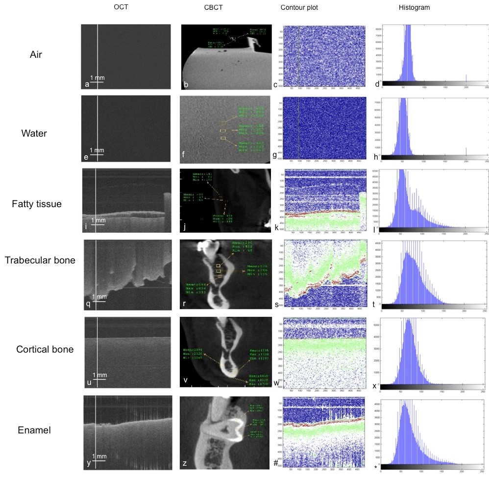
OCT and the corresponding CBCT
image, contour plot for the OCT image, and histogram of the OCT image for air,
water, fatty tissue, trabecular bone, cortical bone, and enamel.
Hassan S. Salehi, PhD, visiting assistant professor of electrical and
computer engineering at the University of Hartford has published a research
article in Elsevier Journal of Oral Surgery, Oral Medicine, Oral Pathology and
Oral Radiology, April 2016. This research work was done in collaboration with
the Stony Brook University School of Dental and the University of Connecticut
(UCONN) School of Dental Medicine.
This paper, "Tissue
characterization using optical coherence tomography and cone beam computed
tomography: A comparative pilot study," reports the imaging of four
types of tissues ex vivo, i.e., human enamel, human cortical bone, human
trabecular bone, fatty tissue plus water and air using optical coherence
tomography (OCT). Furthermore, a method for qualitative and quantitative
analysis of the human specimens was developed utilizing image processing
techniques. The same types of tissues were also imaged using cone beam computed
tomography (CBCT) and grayscale values were measured. The qualitative indices
(intensity profile, contour plot and histogram) for OCT images were able to
provide information regarding surface characteristics as well as changes in
tissue properties at different interfaces. The quantitative index (pixel
intensity values) was also able to render information regarding the distribution
and density of the pixels in different samples. A similar pattern was observed
in the pixel intensity values and grayscale values in both imaging modalities.
Within the limitations of this ex vivo pilot study, it was concluded that OCT
can reliably differentiate between a range of hard and soft tissues.
No comments:
Post a Comment